Inside Unhatched Egg Of Dinosaur That Lived In Argentina 80 Million Yrs Ago
Sauropods — large, four-legged, long-necked dinosaurs, were born with a horn and binocular vision that disappeared as they matured, a study has found.
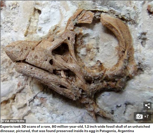
Experts took 3D scans of a rare, 80 million-year-old, 1.2 inch wide fossil skull of an unhatched dinosaur that was found preserved inside its egg in Patagonia, Argentina.
The team think that — had it actually hatched — the juvenile would have used its horn to help it crack its way out of its egg's shell, or for defence or foraging.
The extraordinary fossil specimen represents the most complete and articulate skull found to date of any titanosaur — which were the world's largest-ever land animals.
The largest of these giant plant-eating behemoths — such as Argentinosaurus and Patagotitan — would have grown to reach around 120 feet long.
A different site in Patagonia — Auca Mahuevo — was where sauropod dinosaur eggs were discovered for the first time, among a large nesting site, some 25 years ago.
The provenance of the scanned egg, however, is not known, as it was illegally exported from Argentina before it came to the attention of experts.
The specimen has now been returned to its country of origin for further study, and is housed in the collection of the Museo Municipal Carmen Funes in Plaza Huincul.
'The preservation of embryonic dinosaurs preserved inside their eggs is extremely rare,' said palaeontologist John Nudds of the University of Manchester.
'Imagine the huge sauropods from Jurassic Park and consider that the tiny skulls of their babies, still inside their eggs, are just a couple of centimetres long.'
'We were able to reconstruct the embryonic skull prior to hatching,' he added.
'The embryos possessed a specialised craniofacial anatomy that precedes the post-natal transformation of the skull in adult sauropods.'
'Part of the skull of these embryonic sauropods was extended into an elongated snout or horn, so that they possessed a peculiarly shaped face.'
To recreate what the sauropod embryo would have looked like, the team used a pioneering technique that involved painstakingly dissolving the egg encasing the unhatched dinosaur using acid.
Following this, the specimen was then 'virtually dissected' with a new scanning technology — called 'synchrotron microtomography' — to reveal the inner structure of the unborn sauropod's bones, teeth and soft tissues.
For example, the team were able to identify tiny teeth preserved deep in the dinosaur's jaw sockets, calcified elements of the creature's brain case and even what appear to be the remains of the temporal muscles that move the jaw.
The researchers also detected evidence that the embryonic dinosaurs absorbed calcium from their eggshell to help their growth before they hatched.
'The specimen studied in our paper represents the first 3D preserved embryonic skull of a sauropod,' said paper author and palaeobiologist Martin Kundrat of Slovakia's Pavol Jozef Šafárik University.
'It is a bit unusual for a fossil to be represented just by a skull," he added.
'The specimen perished before completing its development. It had undergone only four-fifths of its incubation period.'
'The most striking feature is head appearance, which implies that hatchlings of giant dinosaurs may differ in where and how they lived in their earliest stages of life.'
'But because it differs in facial anatomy and size from the sauropod embryos of Auca Mahuevo, we cannot rule out that it may represent a new titanosaurian dinosaur.'
With their initial study complete, the team are now hoping to use their technique to examine other embryonic dinosaurs — including other specimens found at the same site in Patagonia.
The full findings of the study were published in the journal Current Biology.


Comments
Post a Comment